I took a very close look at Figure 10 of the Harrit paper - the XEDS-map of 5 elements along with a BSE image of the same region of one chip. My goal originally was to make it more visually clear which pigments are Al-Si-oxides and which are Fe-oxides (that indeed Al+Si map with greyish platelets and Fe with whitish grains), and to my surprise I think I stumbled on two crystal structures that seem to be neither Al-Si nor Fe, but contain elements heavier than Si.
I propose that I found the so-far elusive Strontium Chromate!
But I now need your help to confirm (or refute) this: I haven't found any good reference on what the crystal structure and typical particel size of Sr-Chromate in paint would be. Plus, if y'all could review critically my work, maybe repeat the image manipulations that I did and tell me if what I did is valid to begin with, or if you converge on similar results.
Here it goes. First, a link to the original Figure 10 (too large to display on this forum):
http://i1088.photobucket.com/albums...terial/ActiveThermiticMaterial_Fig10_orig.jpg
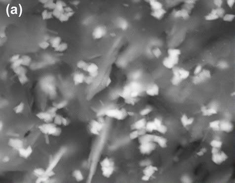
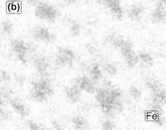
Fig 10a is a BSE image of a region in the the red layer of chip (a), showing an area of about 5µm by 3,5µm. In BSE imaging, the brightness of an obhect depends on the atomic number or mass of its constituent elements. Very light elements such as hydrogen don't show at all (black), light elements such as C or O (atomic numbers 6 and 8) may be dark grey or also be close to invisible, medium elements such as Al or Si (atomic numbers 13 and 14) appear greyish, heavier elements such as Cr, Fe and Sr (atomic numbers 24, 26 and 38) would appear whitish.
Fig 10a:
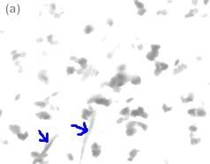
Now, all six parts of Fig 10 contain many shades of gray (or whatever color they are colored in), and it is easy for the human eye to be fooled by varying contrasts and, conversely, difficult to identify areas of equal grey intensity. In other words, it is hard to tell where "greyish" ends and "whitish" begins. I figured a remedy would be to adjust brightness and contrast of the images such that areas below or above a certain intensity would vanish into pure white (or black), so I could see only those area with a minimum signal intensity. I decides to first convert the five colored images to grayscale, then to invert them all, as I am more used to looking at white as a neutral background. Then I increased the brightness of the images, and sometimes played with contrast also, such that areas already close to white would fade to 100% white while areas with a sufficiently string signal stood out clearly as darker shades of gray.
And this is the result for 10a:
Most of the particles are rathe "compact", their lenght:width ratio hardly exceeds 2:1, with those that are longer usually showing a waist or other discontinuites that seem to indicate they are really two particles lumped together. But two structures stand out as being long, thin and straight. Previously, I had seen them as greyish platelets (kaolinite) seen edge-on, but all the other kaolinite platelets, including some that are shown edge-on, have disappeared after my image adjustment - they were simply a darker shade of grey than these two.
So next I tried to see if these "needles" map with Al or Si, or maybe Fe, by looking at the three element maps next to the altered BSE image:
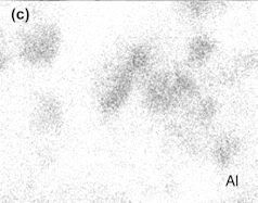
1. BSE and Al:
No match
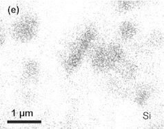
2. BSE and Si:
No match
3. BSE and Fe:
Fe matches the compact grains, but not the "needles"
On the other hand, Al and Si match each other wonderfully:
It is not easy to make the greyish kaolinite stand out, but I tried by increasing contrast greatly and adjusting gamma only slightly. In the adjusted BSE, take both white and black as neutral background of other stuff and look at the grey areas, when you compare with the distribution of Al and Si:
It is my contention that the two needles show too little signal for Si and Al, compared to the other platelets, and are thus not kaolinite. They are not Hematite, either.
The only other possibility I see for our LaClede paint is Strontium Chromate.
Big question: Does Strontium Chromate form "needles" like this?
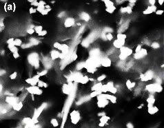
I have one more image (direct link again because of size) that may show Sr-Chromate pigments - simply because these particles are larger than, and more slender, than the many hematite grains - I labelled them with the letter C:
http://i1088.photobucket.com/albums...ede/ActiveThermiticMaterial_Fig05-labeled.jpg
# oysteinbookmark


")