Bob Blaylock
Forklift Operator
When my father passed away in December, I inherited, among other things, his microscope. Lately, I've been going out and collecting bits of gutter water, moss, lichen, and such; mixing it together, and then looking at random drops of it through this microscope.
Just last night, I felt inspired to dig out my cheap digital camera, point it into the microscope's eyepiece, and see what kind of pictures I could get. My expectations were very low. I figured that to get decent pictures though a microscope would probably require more specialized equipment than this.
I was rather pleasantly surprised with the results that I got. Though one could wish for better pictures than these, I've been getting results much better than I thought I could get using such crude methods.
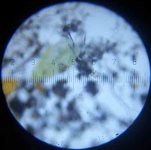
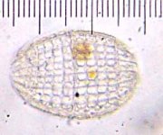
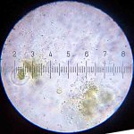
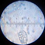
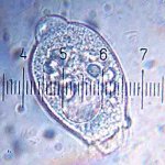
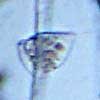
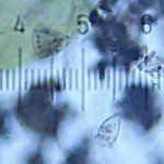
All of these pictures were taken with a Kodak DC3200 camera, using the 15X eyepiece and the 10X objective on the microscope. (The microscope has 5X, 10X and 60X objectives, and 5X, 10X, and 15X eyepieces. The 15X eyepiece has a scale built into it, part of which is visible in the last of the attached pictures here.
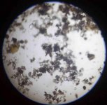
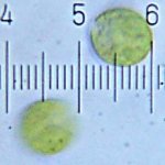
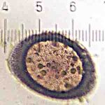
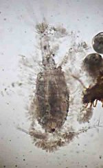
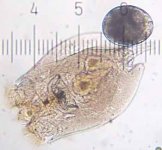
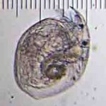
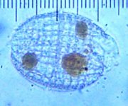
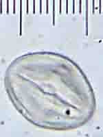
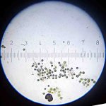
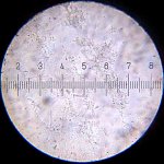
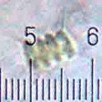
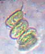
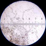
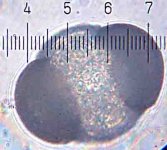
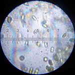
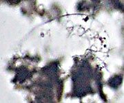
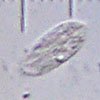
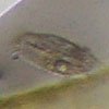
Of the attached pictures, the first and third are of paramecia (not the same one in each picture). I think the second is a dead tartigrade or “water bear”. It wasn't moving. The last two, I think, are of some sort of rotifers.
All of these images are to the same scale. According to notes in my father's hand, written on the inside of the microscope's case, with the 10X objective, each ten marks (that's the small marks, ten from one number to the next) on the scale in the 15X eyepiece is equal to 122 micrometers. That's about a hundred pixels on each of these images, so each pixel on these images is 1.22 micrometers.
My display has about 100 pixels per inch, so each inch on my display is approximately 122 micrometers in the scale of these images.
So these pictures, as they appear on my screen, are about 208X actual size.
Just last night, I felt inspired to dig out my cheap digital camera, point it into the microscope's eyepiece, and see what kind of pictures I could get. My expectations were very low. I figured that to get decent pictures though a microscope would probably require more specialized equipment than this.
I was rather pleasantly surprised with the results that I got. Though one could wish for better pictures than these, I've been getting results much better than I thought I could get using such crude methods.
All of these pictures were taken with a Kodak DC3200 camera, using the 15X eyepiece and the 10X objective on the microscope. (The microscope has 5X, 10X and 60X objectives, and 5X, 10X, and 15X eyepieces. The 15X eyepiece has a scale built into it, part of which is visible in the last of the attached pictures here.
Of the attached pictures, the first and third are of paramecia (not the same one in each picture). I think the second is a dead tartigrade or “water bear”. It wasn't moving. The last two, I think, are of some sort of rotifers.
All of these images are to the same scale. According to notes in my father's hand, written on the inside of the microscope's case, with the 10X objective, each ten marks (that's the small marks, ten from one number to the next) on the scale in the 15X eyepiece is equal to 122 micrometers. That's about a hundred pixels on each of these images, so each pixel on these images is 1.22 micrometers.
My display has about 100 pixels per inch, so each inch on my display is approximately 122 micrometers in the scale of these images.
1_in
1_mm CONVERT
.122_mm
÷
1_mm CONVERT
.122_mm
÷
So these pictures, as they appear on my screen, are about 208X actual size.
Attachments
Last edited:
")