Cont: JFK Conspiracy Theories V: Five for Fighting

Status
Not open for further replies.
So what's the percentage of headshots that occur on camera verses headshots where there are few or no witnesses (other than the killer)?

Just asking for my police friends to make their job easier in case there's some giant visual archive of murder they don't know about.

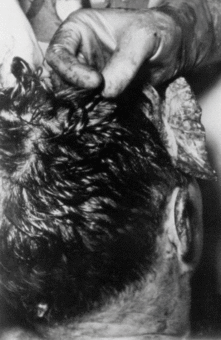
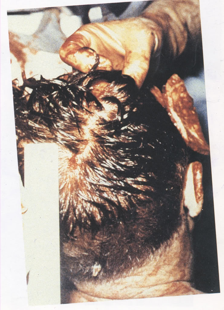
The great-but-sad thing about the United States is our collective knowledge of gunshot wounds to almost every part of the human body.

You have no idea how close that hits to home for me.

I'm at the point where I realize that I should have been studying pentatonic scales instead of ballistic tables when I was a kid.
 
The Warren Commission endorsed the EOP location for the entrance wound in the back of the head. The "cowlick" location for the entrance wound in the back of the head was developed by the Clark panel and the HSCA. The wide difference between the two locations was clearly addressed for the first time in Dr. Finck's 1969 testimony at the trial of Clay Shaw, in which he went on the record disagreeing with the Clark panel's upper "cowlick" placement of the entrance wound.

The only "Clark" mentioned was Dr. Kemp Clark. Here:
Q: Can you tell me, Colonel, whether or not you had at your disposal any information from Dr. Kemp Clark?
MR. DYMOND: If the Court please, we have not been objecting to hearsay but at this point any information of this type would be hearsay unless this doctor spoke with that person and even then it would still be hearsay.
MR. OSER: I didn't ask what the content was, I asked him if he had any information available from Dr. Kemp Clark.
THE COURT: He can say yes or no. Did you understand?
THE WITNESS: There was a Dr. Clark mentioned. I did not talk to him.


The Clark Panel is named after Ramsey Clark, the then-Attorney General of the U.S. Finck did NOT go on the record disagreeing with anything in that Panel's findings. Your claim is false. It is your *interpretation* that he disagreed with the Panel's conclusions, but it is your *interpretation* only. Finck never said that.

https://archive.org/stream/nsia-Aut...nelReport1968/Autopsy Clark Panel 03_djvu.txt

He said there was one bullet entry wound in the head and one bullet exit wound in the head. He said the entry was in the back of the head and the exit was in the right front.

http://mcadams.posc.mu.edu/russ/testimony/finckshaw.htm
Q: Now, Doctor, will you describe to the Jury the nature of this wound which you found on President Kennedy's head and the location of, which you have pointed out on my head?
A: This wound had slightly irregular edges in contrast to the first wound I described in the back of the neck, and I would like to explain at this time the reason for that. The tissue underlying the skin, I have described in the back of the neck is soft tissue, and when the bullet strikes the skin in such an area it does not meet the resistance it meets when there is bone underneath, and this explains the difference in character of those two wounds of entry. The wound in the back of the head showed irregular edges because there was bone close to the scalp corresponding to that scalp wound in the back of the head I just demonstrated, there was a hole in the bone, in the skull of President Kennedy, and I examined it, that hole, from outside the skull and from inside the skull. When examining from outside the skull, I did not see a crater, I saw a hole but there was no crater around it. When I looked at that wound from inside the skull, I saw a definite crater, C-R-A-T-E-R, and this is a certain factor to identify positively the direction of a projectile going through a flat bone such as the skull. To take a practical example, I have seen similar craters in wood, when a bullet goes through and through a pane of wood, and in glass, and it is the difference of the examination between the outer surface and the inner surface that allows the examiner to determine the direction of the bullet. Police officers do that all the time when they examine panes of wood or panes of glass, and I have done so myself. It is an accepted fact.
...
Q: Dr. Finck, in addition to what you have described as a bullet hole of entrance, the location of which you have indicated on my head, would you describe any other damage which you might have found to the skull of the late President Kennedy?
A: In addition to the wound of entry I have discussed in the back of the head, there was a very large wound, irregular, star-shaped, what we call stellate, s-t-e-l-l-a-t-e, approximately five inches in diameter. It was 13 centimeters in diameter, which is approximately five inches and one-eighth. During the course of the autopsy we received from Dallas portions of bone which have the same appearance as the general appearance of the remaining skull of President Kennedy, and on one of the fragments which we could match inside this wound, approximately five inches in diameter, occupying the right side and the top of the head of the President, I saw the beveling I described to you. First identified the outer aspect of the specimen and the inner aspect, I-n-n-e-r, of the specimen to orient the specimen in relation to the wound. After having oriented the specimen as far as the outer and inner surfaces are concerned, I saw this crater when the specimen was viewed from outside which identifies a portion of the wound of exit. You realize that when you have a bullet going through a head at high velocity, the wound of entry may be entire, complete, as in this case, but because of the shattering, s-h-a-t-t-e-r-i-n-g, shattering and explosive force produced by that bullet, the wound of exit is very irregular and very often you don't have all the portions of bone to make a complete skull, some portions are missing, so you cannot do what you do with a complete puzzle, to take the complete pieces and make a complete image. In that case the fragments were matching the wound in a general way, and I could make a positive determination of a wound of exit, of a portion of a wound of exit, in a bone fragment submitted to us during the course of the autopsy, and it was, I would say, between approximately 11:00 o'clock at night -- I can give you the time, it was during the course of the autopsy this fragment was brought to us and allowed us to determine that this was the wound of exit.
Q: Approximately 11:00 o'clock on what date?
A: On the 22nd of November, 1963, the date of the assassination.
Q: Now, Doctor, were any skull fragments delivered to you which were incompatible with your opinion as to the exit area having been on the side of the head?
A: There were none.
Q: There were none. Now, having examined the skull particle which you have testified contained evidence as to which direction the bullet was traveling, and as an expert in the field of Pathology, do you have a definite opinion as to whether the projectile which caused the bone damage exhibited by that particle entered from the front or from the back?
A: I have a definite opinion. I would like to add that that bony specimen brought to us was X-rayed and contained metallic fragments which corroborates the finding of metallic fragments seen at the time of the autopsy on the X-ray film of the head of the President, and the X-ray film was taken before the autopsy of the head, I saw the X-ray film, there were metallic fragments on the X-ray, there were metallic fragments in that bony fragment brought to us during the course of the autopsy, and I have a firm opinion that the bullet entered in the back of the head and exited on the right side of the top of the head producing a very large wound.
Q: Doctor, did you find any evidence which would indicate that the President was hit by more than one shot in the head?
A: No.


You don't get to cite the good doctor as agreeing with you when he destroys your argument for two shots to the head entirely.

He said there was one bullet entry wound in the head and one bullet exit wound in the head. He said the entry was in the back of the head and the exit was in the right front.

You don't get to re-argue the same points you argued and lost six months or more ago. No fringe reset for you.

Hank
 
Last edited:
These are the drawings made under the supervision of Dr. Humes as an accurate representation of the small head wound:

[qimg]https://i.imgur.com/cM8BeTz.png[/qimg]

It appears near the EOP. The Warren Commission endorsed the EOP wound. The existence of the EOP wound is a separate matter than the existence of more than one gunshot to Kennedy's head. The Warren Commission obviously endorsed only one gunshot to the head.

I have searched and not found the date of this drawing that Humes supervised, could you cite a link to the date?
 
I did find the information about the illustrator, Harold Ryberg. The illustrations were completed with only verbal instructions by Humes and Boswell in Mar 1964. They would become exhibits in the WC.

The location of the lower wound in the drawing is above the shoulder line, but the autopsy photograph clearly shows that it is in fact below.
It seems to me that there are communication issues between the doctors and illustrator on placement of the entry wounds. For this reason the illustration representing the head entry wound location would be called into question as to the "exact" position on the head. It is merely a general location and taken with a grain of salt. MJ, you lose again, no conspiracy one head shot in the general area of the EOP. Here is the link that I found, from CT nut berg Barry Keane on a site by Greg Burnham.
http://assassinationofjfk.net/for-the-sake-of-historical-accuracy/
 
You (or some other CT monger) used the term "backspatter" because you or they didn't know what it means and had no idea that somebody else did know what the tern means and would call you out on your ignorance.

Par for the course with you in this thread.

Aristotle and Plato would not approve.

At some point even a train wreck comes to a stop, but you seem to be a perpetual motion machine of fail.

Backspatter is the matter that spurts out of the point of entry for a projectile. It can be identified through crime scene analysis or, if available, video footage of gunshot wounding incidents. I don't know what kind of game you're playing where you try (and fail) to confuse others to intimidate them on a little internet forum.
 
The only "Clark" mentioned was Dr. Kemp Clark. Here:
Q: Can you tell me, Colonel, whether or not you had at your disposal any information from Dr. Kemp Clark?
MR. DYMOND: If the Court please, we have not been objecting to hearsay but at this point any information of this type would be hearsay unless this doctor spoke with that person and even then it would still be hearsay.
MR. OSER: I didn't ask what the content was, I asked him if he had any information available from Dr. Kemp Clark.
THE COURT: He can say yes or no. Did you understand?
THE WITNESS: There was a Dr. Clark mentioned. I did not talk to him.


The Clark Panel is named after Ramsey Clark, the then-Attorney General of the U.S. Finck did NOT go on the record disagreeing with anything in that Panel's findings. Your claim is false. It is your *interpretation* that he disagreed with the Panel's conclusions, but it is your *interpretation* only. Finck never said that.

https://archive.org/stream/nsia-Aut...nelReport1968/Autopsy Clark Panel 03_djvu.txt

He said there was one bullet entry wound in the head and one bullet exit wound in the head. He said the entry was in the back of the head and the exit was in the right front.

http://mcadams.posc.mu.edu/russ/testimony/finckshaw.htm

You don't get to cite the good doctor as agreeing with you when he destroys your argument for two shots to the head entirely.

He said there was one bullet entry wound in the head and one bullet exit wound in the head. He said the entry was in the back of the head and the exit was in the right front.

You don't get to re-argue the same points you argued and lost six months or more ago. No fringe reset for you.

Hank

Hank, what is your problem with lying about obvious things in an effort to confuse other people? Do you think I or other people cannot read? Or maybe you didn't read the testimony.

Q: Colonel, could you tell me, using myself as an example, approximately what the loca- tion in my head would be 100 millimeters above my external occipital protuberance?

A: 100 millimeters is approximately 4 inches. This is the external occipital protuberance. My finger is approximately 4 inches and at a place here which is approximately the location here.

Q: About right here, Colonel, 'cause I can't see you.

A: Approximately here, Mr. Oser.

Q: Now, Colonel, I believe you said that you are familiar with the report of Drs. Carns, Fisher, Morgan, and Moritz, as having reviewed and returned in 1968, I ask you whether or not you disagree with their findings, Colonel, that after viewing the X-rays of the President they found a hole in the President's head 100 millimeters above the occipital protuberance?

A: I can't say I agree or disagree with this for the following reasons: This measurement refers to X-ray films. On of this Panel Review -- what is the exhibit number of this?

Q: I now mark it as State-73 -- 72, I am sorry.

A: On of this Panel Review of 1968, which I read for the first time in 1969, I read: "One of the lateral films of the skull" -- and this refers to a general section heading you will find on "Examination of X-ray Films" on Page 9, as I read this, I interpret this statement of as a measurement based on X-ray films. So there was a difference between measurements made on X-ray films and photographs or photograph ual measurements on the cadaver.

Q: Do you disagree with the fact that these four doctors are qualified in the field of Pathology?

A: They are definitely, three of them, three of them are qualified pathologists, and the fourth doctor is a radiologist.

Q: Radiology is in what field of medicine?

A: Radiology is the study of X-rays for diagnostic reasons or for the reasons of treating with radiation.

Q: Would you say, Colonel, that a radiologist is the best qualified person in the field of medicine to read an X-ray?

A: Yes.

Q: Did you find in reading that report any mention by these four gentlemen, or these four doctors, of any hole in the President's head being one inch slightly above the occipital protuberance bone?

A: I do not find the measurement as one inch to the right of the external occipital protuberance in this State-72.

Q: Colonel, could you step down, and using State Exhibit 70, show me the approximate location in correlation to the size of the diagram, or the illustration, where 100 millimeters would be above the occipital protuberance bone.

A: On which one?

Q: I will repeat my question. Using State Exhibit 70, Colonel, would you show me the approximate location of 100 millimeters above the occipital protuberance bone in relation to the size of this particular illustration as it appears in this exhibit.

MR. DYMOND: If the Court please, this exhibit does not purport to be a scale exhibit and as I said before, it is not a three- dimensional photograph. I doubt if the Doctor could locate this bone, and if he could, any estimate of distance would be useless because it does not purport to be to scale.

MR. OSER: If the Court please, the Doctor used this exhibit saying this is the approximate location he found, and I am now asking him the approximate location that four doctors examining X-rays said it was 100 millimeters above the occipital protuberance bone, and I think he can tell the approximate location of that.

THE COURT: Mr. Dymond's objection is that it is not a picture of the rear of the base of the skull, and for that one reason Mr. Dymond doesn't see how the witness could put it any relation with respect to the rear of the skull and moving laterally across the skull.

MR. DYMOND: He has already done this on Mr. Oser's head, which is three dimensional.

MR. OSER: Still and all he used this exhibit showing at least a portion of the back of the skull and a line going over the top of the skull which would indicate at least to me the approximate mid-part of the head, and I fail to see why the Colonel cannot indicate the approximate location 100 millimeters above the occipital protuberance bone. I know it is not drawn to scale, but I am only asking him for the approximate location.

THE COURT: Could he not do it better in the figure in your autopsy sheet there?

MR. OSER: But, Your Honor, that may well be, but since the Doctor has used this exhibit and said this is where he found a hole, I think the State has a right also to show as a result of the testimony where approximately 100 millimeters was.

THE COURT: You understand the question?

THE WITNESS: Yes, I do, but I can't see how I can be asked to place a wound that was measured on X-rays, I don't understand how I can be asked to put on a illustrative drawing showing the location of the wound as we approximately saw it and not based on measurements on X-rays. Those 100 millimeters --

BY MR. OSER:
Q: Tell me how did the illustrator do it if he didn't have the X-rays and photographs?

A: He did not.

Q: Then how did he do it?

A: Because he was told by Dr. Humes about the approximate location of that wound in the back of the head on the right side and approximately one inch from the external occipital protuberance and slightly above it.

Q: He was told by Commander Humes that?

A: To my knowledge the illustrator making those drawings made them according to the data provided by Dr. Humes.

Q: Let me ask you this then, Colonel: Am I correct in stating that you said that the area I am pointing to right now is the approximate location where four inches above my protuberance bone is?


A: On your head I agree but the measurement of 100 millimeters was made on an X-ray and that is why I am reluctant to say.

Q: Made by a radiologist, one was a member of the American Board of Radiology?

A: I don't know that. That report is signed by four people, there were four to sign it.

Q: Didn't you say one was a radiologist?

A: To my knowledge.

Q: And a radiologist deals in X-rays?

A: A radiologist deals with X-rays and the interpretation of them.

MR. OSER: Again I call for the witness to put the approximate location because there has been testimony on direct examination as well as cross-examination, and because the Defense introduced a picture of Exhibit 388 in Defense Exhibit 67 and I think the State has a right to use this for further witnesses and further cross-examination of the Doctor. I call for this location.

MR. DYMOND: The Doctor has said that he can't do it.

THE COURT: He already testified that the or that there is somewhat of a difference between locations on there and in X-rays and I am not going to force him to do it.

MR. OSER: Then I ask that he mark it on State-68.

THE COURT: If he can do it.

MR. OSER: Four inches above the external occipital protuberance on the descriptive sheet, State-68, and I, this is the Autopsy Descriptive Sheet, and I presume you have used it before for autopsies and I ask that it be so marked there.

THE COURT: If the Doctor can do it.

THE WITNESS: I don't think I can put a wound on a drawing whereas the distance of that wound on an X-ray was given as 100 millimeters I can't do that on something that is different.

MR. OSER: Your Honor, may I ask the witness --

THE COURT: Let's see if I can clarify it. Dr. Finck, on the drawing of the rear of a human being, male, can you place with some kind of a pen or what have you the correction, if one was made, as a result of the four-man panel, as to what you all originally determined. If you can do it and if you can't, you can't do it.

MR. DYMOND: If the Court please, may I submit the Doctor is trying to explain that the distances --

MR. OSER: I don't want Mr. Dymond to testify.

MR. DYMOND: This is in support of my objection.

THE COURT: I will listen.

MR. DYMOND: That the distances on an X-ray measurement is not compatible at all with the distances on this drawing and would be impossible to transpose.

THE COURT: I will accept that. Take the witness stand.

BY MR. OSER:
Q: Doctor, you are familiar with an autopsy descriptive sheet, have you seen something similar to this before and have you ever used something like this before in an autopsy?

A: It is quite common to use worksheets in autopsies.

Q: I ask you again, that wasn't my question, have you used them before?

A: I have used worksheets in autopsies.

Q: And you are telling the Court that you can't mark 100 millimeters above the occipital protuberance bone on that descriptive sheet that you have used before?

MR. DYMOND: If the Court please, it is repetitious. Your Honor has ruled on the question.

THE COURT: I will let the Doctor answer one more time. The question is -- Please read it, Mr. Reporter.

THE REPORTER: Question: "And you are telling the Court that you can't mark 100 millimeters above the occipital protuberance bone on that descriptive sheet that you say you have used before?"

MR. OSER: What is your answer?

THE WITNESS: I could place a wound higher on that drawing but again I don't understand why I am asked to do that.

MR. OSER: I don't think it is for the witness to determine that.

MR. WEGMANN: Let the witness answer.

THE COURT: If you say you can place it, I suggest you leave the witness stand, step down and go place it.

THE WITNESS: That would not be placed on X-rays, that would be a wound higher and approximately in this location.

MR. OSER: These are approximate and we can cover the matter.

BY MR. OSER:
Q: Initial that, please. Thank you, Doctor.

THE WITNESS: Your Honor, at this time I would like to make a comment for the record.

THE COURT: No, sir, you are not running the show. You either answer the question and give an explanation and don't comment.

MR. DYMOND: May we see whether this comment is in the form of an explanation of his answer, Your Honor.

THE COURT: Is the statement that you wish to make in further explanation of your answer to this question?

THE WITNESS: Definitely.

THE COURT: You may do so.

THE WITNESS: The mark I have made --

THE COURT: You can't volunteer information just because you wish to tell us about it. You can only give us answers to a question and then an explanation. There is a difference from what you want to volunteer and what you want to explain. If you want to explain you may do it but you can't volunteer a comment and that is the legal situation of the Court. If this is in further explanation, then I will permit it.

THE WITNESS: The mark I just made on -- what is the exhibit number?

MR. OSER: 68.

THE WITNESS: On Exhibit 68 does not correspond to the wound I have seen at the time of the autopsy. The wound as seen at the time of the autopsy was not as high as that. I did so because repeatedly I am asked to show on this drawing what would the position be of a wound approximately four inches or 100 millimeters above the external occipital protuberance, but I don't endorse the 100 millimeters for this drawing. Again the measurement was made on X-rays. I was more or less forced to put that on this exhibit.

MR. OSER: I want the record to reflect the witness was not forced.

THE WITNESS: I was asked to show on this drawing a wound four inches from the external occipital protuberance.

THE COURT: Let's go on to another area.


...

Q: Now, Doctor, referring to State Exhibit-68, and more particularly the sketch on the lower portion of this, and the red dot which you placed on the right-hand figure of that sketch, does that purport to represent accurately the location of the back head wound as described in the reviewing pathological report of 1968?

A: It does not, and let me explain this. I was asked yesterday by Mr. Oser to place a wound 4 inches or 100 millimeters, approximately, above the external occipital protuberance. The reason for doing so was that in the 1968 panel, P-A-N-E-L, in the chapter entitled "X-rays," this is S-72 on , you will find this figure of 100 millimeters above the external occipital protuberance, but in the first line of that paragraph you see the word "films" on one of the lateral films of the skull, a hole measuring approximately 8 millimeters in diameter on the outer surface of the skull and as much as 20 millimeters on the external surface can be seen in profile approximately 100 millimeters above the external occipital protuberance, so this measurement of 100 millimeters or 4 inches refers to a measurement made on X-ray film and not on the photographs or skull itself. I saw that wound of entry in the back of the head at approximately 1 inch or 25 millimeters to the right and slightly above the external occipital protuberance, and it was definitely not 4 inches or 100 millimeters above it, so I was asked to put on the drawing a measurement coming from the X-ray measurement.


...

RE-CROSS EXAMINATION BY MR. OSER:
Q: Colonel, in referring to State Exhibit-68, the autopsy descriptive sheet, can you tell me whether or not the mark placed on the rear portion or the rear diagram of a body which is indicated with the arrow and marked ragged, slating 15 x 6 millimeter, can you tell me whether or not this spot on this diagram corresponds to a position on the head of 1 inch, approximately 1 inches above the external occipital protuberand or does it apply to 100 millimeters above the external occipital protuberance?

A: It refers to an approximate location on this drawing and it refers to the wound I saw at 1 inch from the external occipital protuberance.

Q: All right.

A: It was definitely not 4 inches or 100 millimeters above it.


Thus, Finck's 1969 testimony at the trial of Clay Shaw marks the first time a JFK autopsy doctor specifically denied the cowlick entry theory.
 
I did find the information about the illustrator, Harold Ryberg. The illustrations were completed with only verbal instructions by Humes and Boswell in Mar 1964. They would become exhibits in the WC.


It seems to me that there are communication issues between the doctors and illustrator on placement of the entry wounds. For this reason the illustration representing the head entry wound location would be called into question as to the "exact" position on the head. It is merely a general location and taken with a grain of salt. MJ, you lose again, no conspiracy one head shot in the general area of the EOP. Here is the link that I found, from CT nut berg Barry Keane on a site by Greg Burnham.
http://assassinationofjfk.net/for-the-sake-of-historical-accuracy/

Humes directed the back wound to be drawn higher as the result of an agenda to endorse a single bullet entering the back and exiting the throat. Here is a 1964 clip of Humes appearing on CBS swearing that the Ryderg drawing's placement of the back wound is accurate: http://www.youtube.com/watch?v=ipwOmCLMpqo&t=27m34s
 
Last edited:
Are those the ONLY images in the WCR?
How close is that sketch to the "cowlick"?
Why is no wound in the "EOP" visible in the photographs?

On the existing BOH photographs, if one of the ~two semicircular dark spots in the hairline are not the EOP wound, then it could be hiding under a bit of hair. There were autopsy photographs that went missing before the official inventory was made. The autopsy doctors and photographer John Stringer have described taking clear photographs of an entry wound near the EOP in the scalp and skull.
 
I did find the information about the illustrator, Harold Ryberg. The illustrations were completed with only verbal instructions by Humes and Boswell in Mar 1964. They would become exhibits in the WC.


It seems to me that there are communication issues between the doctors and illustrator on placement of the entry wounds. For this reason the illustration representing the head entry wound location would be called into question as to the "exact" position on the head. It is merely a general location and taken with a grain of salt. MJ, you lose again, no conspiracy one head shot in the general area of the EOP. Here is the link that I found, from CT nut berg Barry Keane on a site by Greg Burnham.
http://assassinationofjfk.net/for-the-sake-of-historical-accuracy/


You're correct, and in fact, in his testimony to the Warren Commission, Humes took care to not only point that out, but also said the drawings were of necessity only schematic in nature, as Rydberg had only his meager verbal description to work from, and did not have any photos or X-rays from which to work.

\http://mcadams.posc.mu.edu/russ/testimony/humes.htm
Commander HUMES - Yes, sir. We had made certain physical measurements of the wounds, and of their position on the body of the late President, and we provided these and supervised directly Mr. Rydberg in making these drawings.
Mr. SPECTER - Have you checked the drawings subsequent to their preparation to verify their accuracy?
Commander HUMES - Yes, sir.
Mr. SPECTER - And proportion?
Commander HUMES - I must state these drawings are in part schematic. The artist had but a brief period of some 2 days to prepare these. He had no photographs from which to work, and had to work under our description, verbal description, of what we had observed.
...
(Later) Commander HUMES - These exhibits again are schematic representations of what we observed at the time of examining the body of the late President.


Hank
 
Last edited:
On the existing BOH photographs, if one of the ~two semicircular dark spots in the hairline are not the EOP wound, then it could be hiding under a bit of hair.
Why are CTists so in love with "could be" and scared to death of "is". Is a second gun shot hiding under the hair other than the clear photo of a "red spot" which is TOTALLY NOT a gun shot entrance wound?

There were autopsy photographs that went missing before the official inventory was made. The autopsy doctors and photographer John Stringer have described taking clear photographs of an entry wound near the EOP in the scalp and skull.
Actual evidence for a second entrance wound?
 
You're correct, and in fact, in his testimony to the Warren Commission, Humes took care to not only point that out, but also said the drawings were of necessity only schematic in nature, as Rydberg had only his meager verbal description to work from, and did not have any photos or X-rays from which to work.

\http://mcadams.posc.mu.edu/russ/testimony/humes.htm
Commander HUMES - Yes, sir. We had made certain physical measurements of the wounds, and of their position on the body of the late President, and we provided these and supervised directly Mr. Rydberg in making these drawings.
Mr. SPECTER - Have you checked the drawings subsequent to their preparation to verify their accuracy?
Commander HUMES - Yes, sir.
Mr. SPECTER - And proportion?
Commander HUMES - I must state these drawings are in part schematic. The artist had but a brief period of some 2 days to prepare these. He had no photographs from which to work, and had to work under our description, verbal description, of what we had observed.
...
(Later) Commander HUMES - These exhibits again are schematic representations of what we observed at the time of examining the body of the late President.


Hank

Why, it's almost as if CTists deliberately misrepresent things dishonestly.
 
Hank, what is your problem with lying about obvious things in an effort to confuse other people? Do you think I or other people cannot read? Or maybe you didn't read the testimony.
[testimony deleted]
Thus, Finck's 1969 testimony at the trial of Clay Shaw marks the first time a JFK autopsy doctor specifically denied the cowlick entry theory.

Does the Rydberg drawing put the entry wound in the head at the level of the top of the ears? YES.

Did the autopsy photos show the same? YES.

Are you arguing for an entry wound well below that, at the extent of the bottom of the hairline? YES.

You lose.

Hank
 
Last edited:
Humes directed the back wound to be drawn higher as the result of an agenda to endorse a single bullet entering the back and exiting the throat. Here is a 1964 clip of Humes appearing on CBS swearing that the Ryderg drawing's placement of the back wound is accurate: http://www.youtube.com/watch?v=ipwOmCLMpqo&t=27m34s

At ~29:59 in the video Humes describes one of the drawings used to describe the wounds locations. He states that the drawings are used to indicate the general area of the entry wound, NOT the precise location.

At ~ 32:59 He states there is conclusive evidence that the bullet entered the head and EXITED the head in the right front of the skull. How much more do you need to debunk your pet theories? Nowhere does he indicate the bullet that entered the back of the head exited the throat.

Further you accuse Hank of not being able to read, how about you? Here is my
post:
...

It seems to me that there are communication issues between the doctors and illustrator on placement of the entry wounds. For this reason the illustration representing the head entry wound location would be called into question as to the "exact" position on the head. It is merely a general location and taken with a grain of salt. MJ, you lose again, no conspiracy one head shot in the general area of the EOP. Here is the link that I found, from CT nut berg Barry Keane on a site by Greg Burnham.
http://assassinationofjfk.net/for-the-sake-of-historical-accuracy/

Notice that I indicated that the drawings could not be used to locate the exact position, not that they were incorrect. I stand by that statement, although there were not communication issues as I poste earlier.
 
Does the Rydberg drawing put the entry wound in the head at the level of the top of the ears? YES.

Did the autopsy photos show the same? YES.

Are you arguing for an entry wound well below that, at the extent of the bottom of the hairline? YES.

You lose.

Hank

The Rydberg drawings are meant to show the wound as measured in the autopsy report, 2.5 centimeters to the right and slightly above the EOP.

The red spot on the BOH photographs appears to be situated about a couple of inches above "the level of the top of the ears". The head is tilted back, so it can be hard to see, but look at this stereoscopic gif of the BOH photographs here:

JFK-Autopsy-Photos-GIF.gif


To me, the red spot has the appearance of being somewhere between the actual EOP and the area 4 inches higher endorsed by some as an entry point in the skull. Maybe the red spot a little more than ~2 inches above the EOP? If you can show me how the BOH photographs can show the red spot as being 4 inches above the EOP, be my guest. So far, there is no actual reason to think the red spot could correlate to the depressed cowlick fracture on the X-rays 4 inches above the EOP.

The bottom of the hairline is significantly below the EOP. The white matter at the bottom of the hairline with the semicircular dark spot below it is below the EOP. The other semicircular dark spot to the right and at the 1 O' clock position, I don't know.

t3eaa68_JFKcolor_boh_autopsy_photo.jpg
 
Last edited:
At ~29:59 in the video Humes describes one of the drawings used to describe the wounds locations. He states that the drawings are used to indicate the general area of the entry wound, NOT the precise location.

Dr. Humes was claiming that the autopsy face sheet diagram only showed a general area of the wounds, then claimed that the Rydberg drawings are a more precise depiction of the wounds.

At ~ 32:59 He states there is conclusive evidence that the bullet entered the head and EXITED the head in the right front of the skull. How much more do you need to debunk your pet theories? Nowhere does he indicate the bullet that entered the back of the head exited the throat.

But does Humes indicate that there existed an entry wound near Kennedy's EOP? Let your logical deductions start with that. Can a 6.5 round enter there and exit the top of the head while staying consistent with all of the official evidence? It doesn't seem like it can.
 
On the existing BOH photographs, if one of the ~two semicircular dark spots in the hairline are not the EOP wound, then it could be hiding under a bit of hair. There were autopsy photographs that went missing before the official inventory was made. The autopsy doctors and photographer John Stringer have described taking clear photographs of an entry wound near the EOP in the scalp and skull.

Furthermore, Tomtomkent can not exclude the theory that the open-cranium photographs do indeed show the EOP wound.

The prime candidate is the dark area in the lower-right corner:

OxNBCsc.gif


See the size of the doctor's fingers and how they are beneath the skull bone in the foreground where the dark spot lays. That would make it a good fit for the elliptical 15x6mm wound described in the autopsy report. In this orientation, it would show the top of the head (occipital-parietal, with the center of the image being the "cowlick" area). His chin would probably have to be resting on his chest to show everything in this orientation.
 
Last edited:
Furthermore, Tomtomkent can not exclude the theory that the open-cranium photographs do indeed show the EOP wound.

The prime candidate is the dark area in the lower-right corner:

[qimg]https://i.imgur.com/OxNBCsc.gif[/qimg]


See the size of the doctor's fingers and how they are beneath the skull bone in the foreground where the dark spot lays. That would make it a good fit for the elliptical 15x6mm wound described in the autopsy report. In this orientation, it would show the top of the head (occipital-parietal, with the center of the image being the "cowlick" area). His chin would probably have to be resting on his chest to show everything in this orientation.

I can exclude that by the obvious virtue that they show no such thing, are cropped and out of context, and clearly you have no skill to know what you are looking at.

No medical record at the time suggests your belief has foundation, and your interpretation of the photographs is of no more importance than you belief a blatant wound is "a red splotch".
 
Backspatter is the matter that spurts out of the point of entry for a projectile. It can be identified through crime scene analysis or, if available, video footage of gunshot wounding incidents. I don't know what kind of game you're playing where you try (and fail) to confuse others to intimidate them on a little internet forum.

Provide an actual cite from a forensic science source (not a CT advocacy site or person) or fold your tent. You used a term (probably copped it from someone else that didn't know what they were talking about) and don't understand what the term means. Using technical terms that sound all sciencey may impress the hell out of other know-nothings, but is guaranteed to bite you on the ass if you encounter individuals that are trained in the subject matter.

My intent is not to intimidate. My intent is to refute absolute nonsense when I encounter it. If you'd take the time to study forensic science in general as it relates to GSW's and ballistics instead of playing this stupid pin-the-headwound jive you'd be much better off.

Are you going to keep digging your hole to nowhere, or try to learn enough about the subject matter in general so you can participate from an educated perspective? And when I say subject matter, I'm not talking about JFK centric dueling CT's - Get yourself some International Wound Ballistics Association papers and get to woodshedding. In case you don't know what that term means:

http://www.urbandictionary.com/define.php?term=woodshedding
 
I can exclude that by the obvious virtue that they show no such thing, are cropped and out of context, and clearly you have no skill to know what you are looking at.

No medical record at the time suggests your belief has foundation, and your interpretation of the photographs is of no more importance than you belief a blatant wound is "a red splotch".

The 1966 inventory of the autopsy photographs, written by Humes et. al, describes the open-cranium photographs as showing a wound of entrance in the posterior skull.
 
Dr. Humes was claiming that the autopsy face sheet diagram only showed a general area of the wounds, then claimed that the Rydberg drawings are a more precise depiction of the wounds.

Not in this video, listen again and point out at what timestamp he stated that.

EDIT2: He does state that the Ryberg drawing if the back wound was intended to be more precise.

But does Humes indicate that there existed an entry wound near Kennedy's EOP? Let your logical deductions start with that. Can a 6.5 round enter there and exit the top of the head while staying consistent with all of the official evidence? It doesn't seem like it can.

If you look at the video he does not state any particular part of the skull, but places his hand at the back of his skull to indicate the location, I'm afraid I don't have x ray vision, as to where his hand was placed.
EDIT:
And you ignored the link that Hank provided:
(Later) Commander HUMES - These exhibits again are schematic representations of what we observed at the time of examining the body of the late President.
 
Last edited:
Status
Not open for further replies.

Back
Top Bottom